
Condition
Endocrinology
Type 2 Diabetes Mellitus
Type 2 Diabetes Mellitus is an established clinical disorder categorized under Endocrinology. Characterized by pathophysiological involvemen...
12 min readRead
Explore health education guides, medicine information, condition resources, interaction checking, and practical health calculators.
Evidence-based disease overviews, clinical symptoms, risk factors, diagnostic criteria, and management options.
Type 2 Diabetes Mellitus is an established clinical disorder categorized under Endocrinology. Characterized by pathophysiological involvemen...
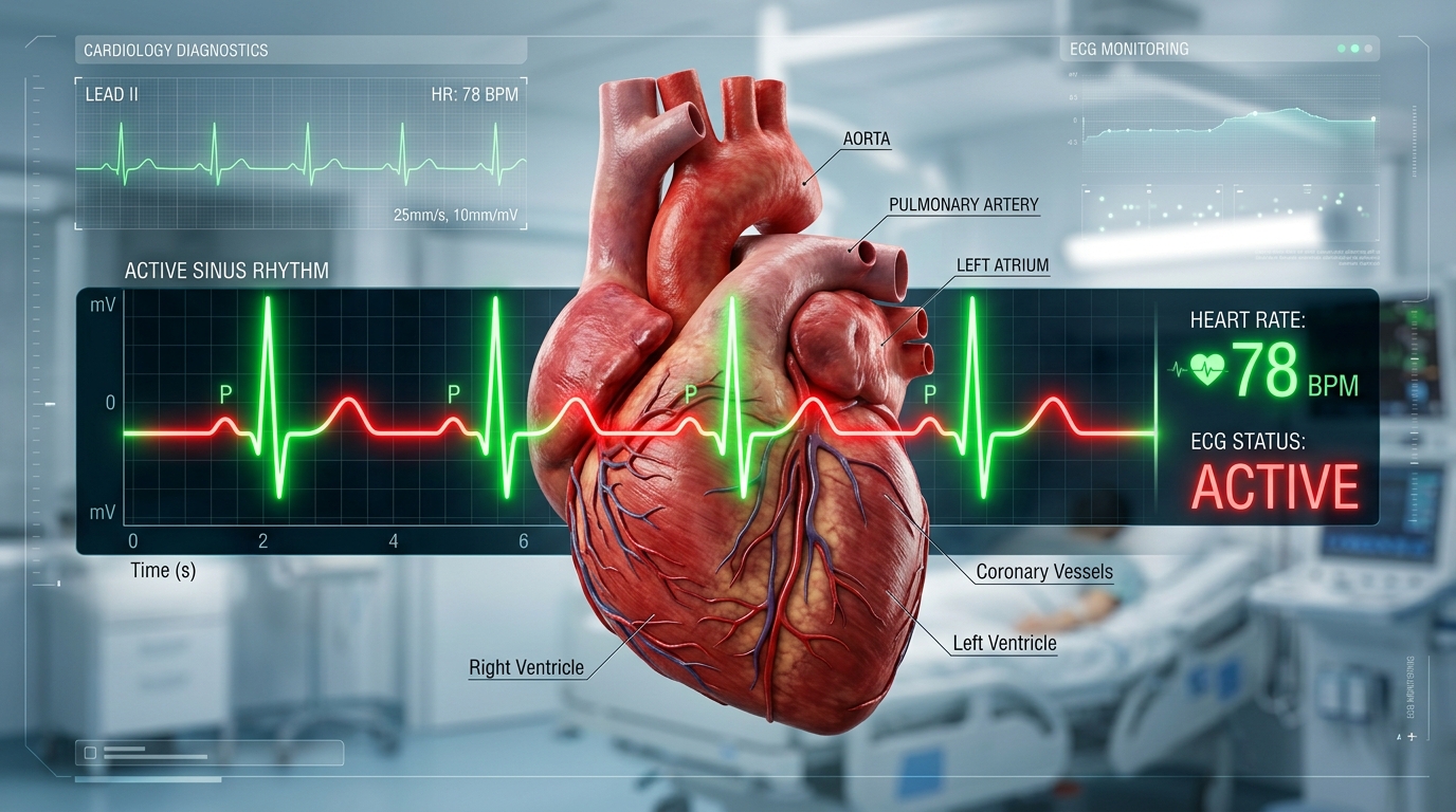
Essential Hypertension is an established clinical disorder categorized under Cardiology. Characterized by pathophysiological involvement of...
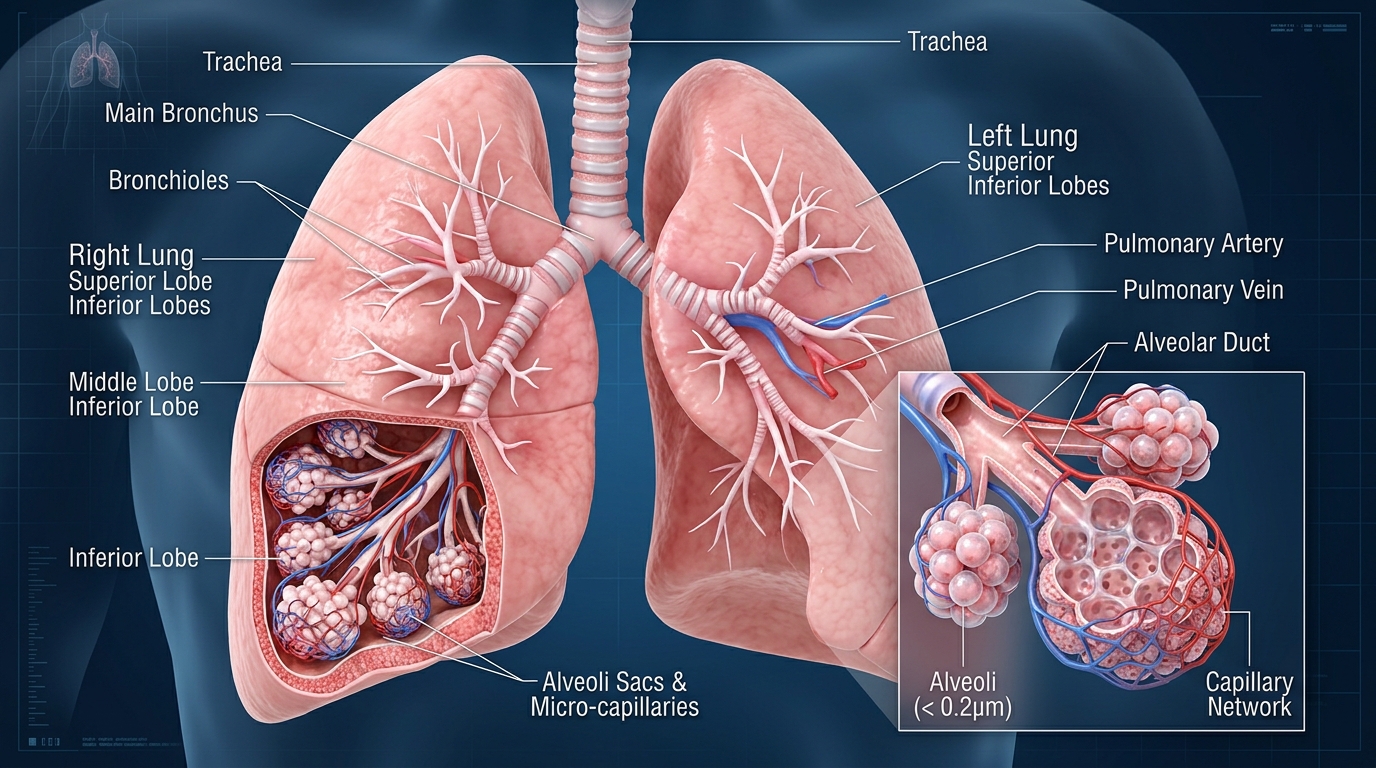
Bronchial Asthma is an established clinical disorder categorized under Pulmonology. Characterized by pathophysiological involvement of the r...

Leukemia is a broad term for malignant cancers of the body's blood-forming tissues, including the bone marrow and the lymphatic system. It i...
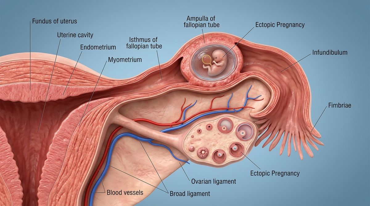
Ectopic pregnancy is a pregnancy in which a fertilized egg implants and begins to develop outside the normal cavity of the uterus (womb). In...

Migraine is an established clinical disorder categorized under Neurology. Characterized by pathophysiological involvement of the central ner...
Browse 16 additional condition guides covering symptoms, diagnosis, and care.
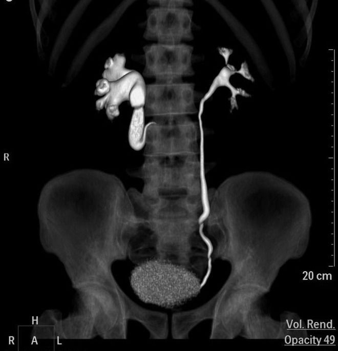
A rare congenital condition in which the right ureter abnormally curves behind the main vein of the abdomen (inferior vena cava), which can squeeze the tube and block urine flow from the kidney.
Retrocaval Ureter (also called Circumcaval Ureter) is an uncommon developmental condition present from birth. Usually, the ureters (tubes carrying urine from kidneys to the bladder) run in front of all major blood vessels. In this anomaly, the right ureter travels behind the Inferior Vena Cava (the body's largest abdominal vein), loops around it, and then returns to its normal path. The pressure between the vein and spine can compress the ureter, causing urine backup and kidney swelling.
Medical Disclaimer: This information is for educational purposes only and is not a substitute for professional medical advice, diagnosis, or treatment.
We investigate popular health claims by separating what people believe from what scientific evidence actually supports.
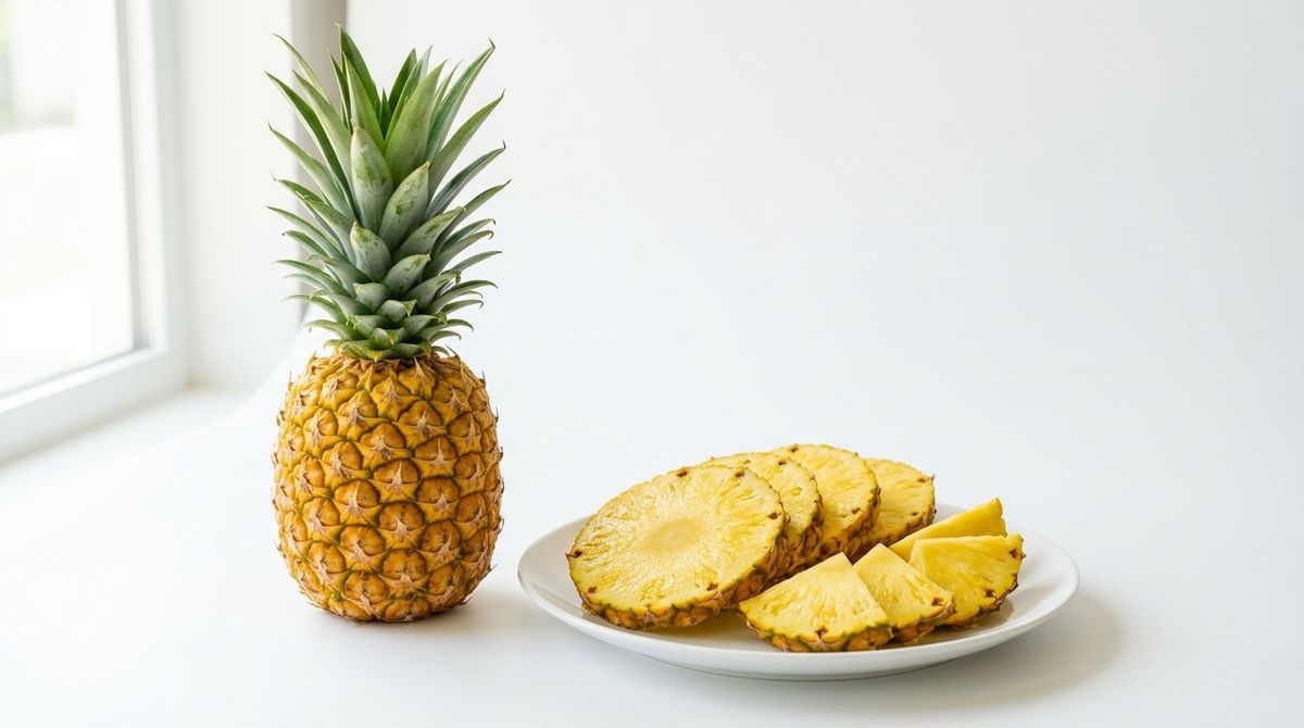
The popular claim investigated — bromelain, mucociliary clearance, and what science actually says.
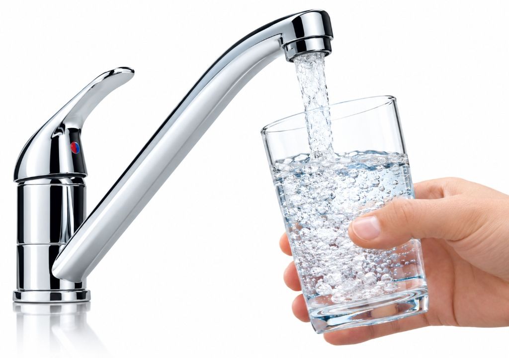
Kidney filtration, ADH hormone, thermoregulation — the real biology of hydration explained.

Vitamin D synthesis, circadian rhythm, UV damage — the complete science of sun exposure.
Yogurt probiotics, detox cleanses, garlic immunity, olive oil, coffee, and coconut water — evidence separated from myth.
Immune defenses naturally decline past 50. Protecting yourself against preventable illnesses preserves vitality and long-term health.
As our bodies mature, waning childhood antibodies and natural immunosenescence leave adults vulnerable to debilitating conditions like shingles, pneumococcal pneumonia, and severe influenza complications.
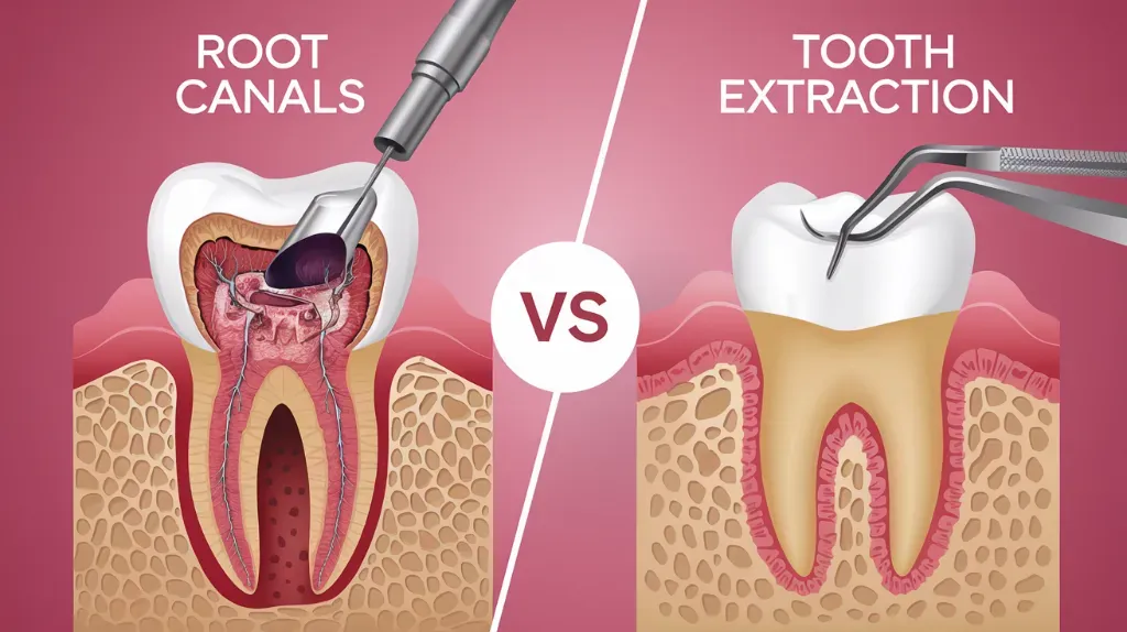
Understand the treatment that can help save a damaged or infected tooth.
Understand the symptoms, procedure, pain, recovery, aftercare, and key differences between root canal treatment and tooth extraction.
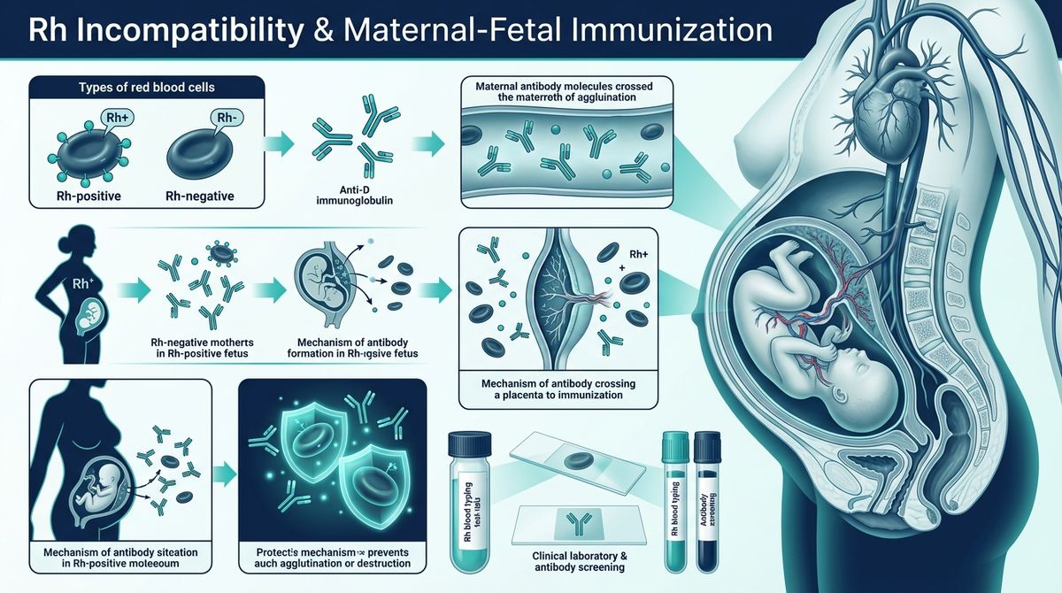
When an Rh-negative mother carries an Rh-positive baby, fetomaternal blood mixing can trigger lifelong antibody production against fetal blood cells. Learn the essential blood tests used to check if antibodies are in the maternal bloodstream and how timely Anti-D Immunoglobulin (RhoGAM) prevents Hemolytic Disease of the Fetus and Newborn (HDFN).
Before giving Anti-D Immunoglobulin (RhoGAM), obstetricians must verify whether the mother has already been sensitized (isoimmunized). If maternal anti-D antibodies have already formed, RhoGAM cannot reverse them. The gold-standard diagnostic tool is the Indirect Coombs Test (Indirect Antiglobulin Test - IAT).
Maternal Antibody Screen / Circulating Anti-D Test
Checks whether the Rh-negative mother has ALREADY developed circulating anti-Rh (anti-D) antibodies against fetal red blood cells.
Serial Antibody Quantitation Assay
Measures the precise concentration/strength of circulating anti-D IgG antibodies in maternal blood via serial dilutions (e.g., 1:2, 1:4, 1:8, 1:16, 1:32).
Blood Group & Rh Typing (Forward & Reverse)
Determines whether the mother is Rh-negative (dd) and verifies the biological father’s Rh status (Rh-positive DD or Dd vs Rh-negative dd).
Newborn Cord Blood Antiglobulin Test
Tests whether maternal anti-D antibodies have already crossed the placenta and physically bound onto the newborn baby’s red blood cells.
Fetomaternal Hemorrhage (FMH) Quantification
Quantifies the exact volume of fetal red blood cells that have crossed into maternal circulation during delivery or traumatic events.
Non-Invasive Prenatal Testing (NIPT) for Fetal RhD
Analyzes circulating fetal DNA fragments in maternal plasma to non-invasively determine whether the unborn baby is RhD-positive or RhD-negative.
Whether you are protecting healthy kidneys or managing Chronic Kidney Disease (CKD Stages 1 through 5), precision nutrition is your most potent clinical defense. Learn how managing the 5 Core Renal Pillars—Sodium, Potassium, Phosphorus, Protein, and Fluids—protects nephrons, prevents toxic uremic buildup, and sustains energy and longevity.

8+ Kidney-Friendly Foods for Renal Diets
Blueberries deliver anthocyanin antioxidants that protect glomerular capillaries without potassium overload. Red bell peppers supply full-spectrum vitamin A, C, and B6 at only 211 mg K+ per 100g. Cauliflower provides the perfect low-potassium mashed potato alternative (176 mg K+). Extra virgin olive oil adds heart-protective oleic acid and polyphenols with zero electrolyte load.

HBV Protein · Zero Phosphorus Yolk · Rich Antioxidants
Two egg whites deliver High Biological Value (HBV) protein with virtually zero phosphorus (all egg phosphorus is in the yolk). Paired with vitamin-C-rich red and yellow bell peppers (boosts iron absorption) and antioxidant-rich blueberries (114 mg K+ / 100g). Drizzled with anti-inflammatory garlic olive oil. Estimated total: ~15g protein, <200mg sodium, ~250mg potassium.
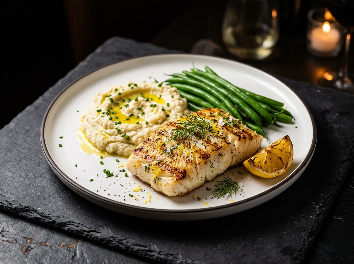
Omega-3 Rich · Low Potassium · Anti-Inflammatory
A 3 oz grilled wild cod fillet delivers 20–24g of high-quality protein with moderate phosphorus (180mg) and heart-protective omega-3 EPA/DHA. Paired with creamy steamed cauliflower mash (176 mg K+/100g) replacing dangerous mashed potatoes, and crisp steamed green beans. Total dinner: ~24g protein, ~50mg sodium, ~480mg potassium — within Stage 3 CKD targets.
Nutrient requirements shift dramatically as glomerular filtration rate (eGFR) changes. Switch below to see precise targets.
Significant reduction in functional nephrons. Accumulation of urea nitrogen, early phosphate retention, impaired vitamin D activation, and secondary hyperparathyroidism.
Protein restriction without protein-energy wasting, monitoring serum bicarbonate (>22 mEq/L) to prevent metabolic acidosis.
Explore clinical safety profiles, electrolyte values, and exact kitchen substitutions to protect failing kidneys while maintaining gourmet flavor.
Rich in vitamin C, folate, and sulforaphane. An outstanding low-potassium alternative that can be mashed to replace high-potassium mashed potatoes.
Packed with anthocyanin antioxidants that fight systemic inflammation and protect glomerular capillaries without potassium overload.
Unlike most vegetables, red bell peppers provide abundant vitamin A, C, and B6 while maintaining an exceptionally low potassium profile.
The gold-standard High Biological Value (HBV) protein. 100% pure protein with almost zero phosphorus, as all egg phosphorus is concentrated in the yolk.
Calorie-dense healthy monounsaturated fat (oleic acid) and polyphenols that combat cardiovascular risk in renal patients without mineral load.
Contains allicin and quercetin. Delivers intense savory flavor so you can eliminate added table salt without compromising taste.
Cruciferous vegetable loaded with vitamins K and C, insoluble fiber, and phytochemicals that promote gut microbiome health and lower uremic toxins.
High in pectin, a soluble fiber that lowers cholesterol, regulates blood glucose, and reduces nitrogenous load.
A single banana delivers roughly 20% of an advanced CKD patient’s entire daily potassium budget. Can trigger sudden cardiac arrhythmia.
Severe hyperkalemia hazard if boiled whole or fried. Can only be eaten in strict moderation IF leached using the clinical 4-step boiling method.
FATAL NEUROTOXIN DANGER. Contains carambolin, an oxalate neurotoxin. Healthy kidneys clear it, but in CKD it crosses into the brain causing intractable hiccups, seizures, and death.
Loaded with inorganic phosphoric acid. Unlike organic plant phosphorus (40% absorbed), inorganic phosphate is 90–100% absorbed into the bloodstream, accelerating vascular calcification.
A triple threat for kidneys: loaded with sodium chloride (fluid retention), inorganic phosphate salts, and advanced glycation end products.
Brands like Nu-Salt or LoSalt substitute sodium with potassium chloride. One pinch can cause severe hyperkalemia and acute fatal cardiac arrest in CKD patients.
Supplies high-quality protein and omega-3 fatty acids (EPA/DHA) that lower systemic triglycerides. Must be portion-controlled (3 to 4 ounces) in Stages 3–4.
Much lower in potassium and phosphorus than cow’s milk (which contains ~380 mg K and 230 mg P per cup). Check label to confirm no added tricalcium or potassium phosphate.
Root vegetables like potatoes, carrots, yams, and squash can be made safer for CKD Stages 3–5 by leaching excess potassium ions into warm water before cooking.
Engineered for CKD Stage 3 (~1,800 kcal, ~50g protein, <1,500mg sodium, low potassium, low phosphorus).
2 egg whites scrambled in olive oil with diced red bell peppers + 1/2 cup fresh blueberries + 1 slice sourdough toast.
1 small crisp apple sliced and dusted with ground cinnamon (zero sodium, rich in soluble pectin fiber).
3 oz fresh cod with lemon dill + 1 cup steamed cauliflower mash drizzled with garlic-infused olive oil + steamed green beans.
3 oz chicken tenderloins sautéed with shredded purple cabbage, garlic, and scallions over 1/2 cup jasmine white rice.
Failing kidneys lose the ability to clear specific dietary toxins and mineral spikes. Avoid these catastrophic hazards:
NEVER eat star fruit or drink star fruit juice. It contains carambolin, an oxalate neurotoxin that accumulates in renal failure, causing seizures, coma, and permanent brain damage.
"Low sodium" salts replace sodium with potassium chloride. Just 1 teaspoon delivers over 800 mg of potassium, risking sudden fatal ventricular tachycardia.
Over-the-counter painkillers (Ibuprofen, Naproxen, Diclofenac) constrict renal afferent arterioles, precipitating acute on chronic renal failure.
Inspect ingredient lists for "Pyrophosphate", "Dicalcium phosphate", or "Phosphoric acid". Inorganic forms are 100% absorbed, petrifying blood vessels.
Every kidney is unique. Serum potassium, eGFR, proteinuria, and bicarbonate fluctuate with medication and disease etiology. Always work alongside a Registered Dietitian Nutritionist (RDN) specializing in clinical nephrology.
Explore how modern evidence-based physical therapy restores physiological kinematics, reverses chronic musculoskeletal pain, avoids invasive surgery, and retrains neurological motor pathways.

Physiotherapy is not merely passive massage or generalized stretching. It is a precise medical discipline grounded in mechanobiology. When therapeutic mechanical loads are applied across muscles, tendons, and articular cartilage, integrin receptors on cell membranes deform. This converts physical stress into biochemical signals (the mechanotransduction pathway), instructing tenocytes to lay down parallel Type I collagen fibers, stimulating chondrocyte proteoglycan synthesis, and remodeling damaged tissue back to high tensile strength.

Hands-on clinical manipulation, oscillatory glide, and myofascial release
Restores physiological joint arthrokinematics (roll, glide, spin), breaks intra-articular adhesions, and relieves localized capsular restriction.
Maitland Grades I–V oscillatory mobilization and Kaltenborn traction stimulate mechanoreceptors (Type I and II Ruffini & Pacinian corpuscles) in joint capsules, inhibiting spinal nociceptive transmission via the Melzack-Wall pain gate while improving synovial fluid circulation.
Click any modality below to inspect clinical protocols and imaging.
Hands-on clinical manipulation, oscillatory glide, and myofascial release
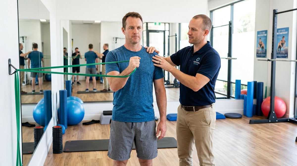
Progressive active loading, eccentric conditioning, and core motor recruitment
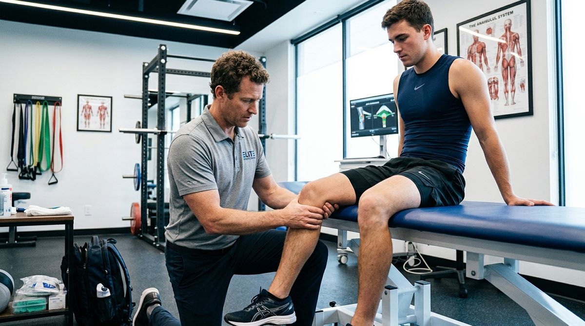
Kinetic chain assessment, plyometrics, and functional performance screening

Harnessing neuroplasticity to restore balance, coordination, and independent ambulation
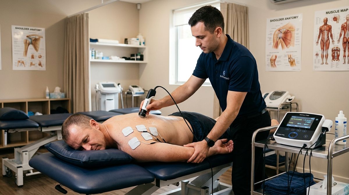
Targeted bioelectric stimulation, ultrasound, and thermal tissue modulation
A rigorous exploration of how active movement therapy remodels human connective tissue, recalibrates central nervous system pain circuits, and out-performs passive pharmacological treatments in long-term musculoskeletal health.
For much of the twentieth century, clinical medicine responded to acute musculoskeletal injuries, severe low back pain, and post-operative joint trauma by prescribing prolonged bed rest, rigid splinting, and immobilization. Today, modern physical medicine and rehabilitation (PM&R) recognizes that prolonged immobilization is among the most biologically destructive interventions a clinician can prescribe to the human locomotor system.
Within 48 to 72 hours of complete bed rest, striated skeletal muscle exhibits rapid protein catabolism, loss of sarcomeres in series, and significant motor unit de-recruitment. Concurrently, synovial fluid flow within joint capsules stagnates, deprivation of cyclic hydrostatic pressure accelerates articular cartilage dehydration, and collagen fibers in healing tendons synthesize in a disorienting, tangled, non-functional meshwork known as cross-linked scar tissue.
Modern physiotherapy establishes an entirely new paradigm: Movement is biological medicine. The human musculoskeletal system is fundamentally adaptive, designed to maintain bone density, ligament tensile strength, and myofascial compliance through the continuous application of controlled, physiological mechanical stresses. Rather than immobilizing an injured limb, physical therapists utilize the principle of Optimal Loading—applying specific vectors of force, tension, and compression to stimulate cellular regeneration while protecting fragile structural repairs from catastrophic reinjury.
The obsolete RICE protocol (Rest, Ice, Compression, Elevation) has been superseded by POLICE: Protection, Optimal Loading, Ice, Compression, Elevation. Complete rest is replaced by early, controlled mechanical loading to stimulate mechanoreceptors, preserve neuromuscular synapses, and prevent arthrogenic muscle inhibition.
At the molecular level, physiotherapy exercises do not just "build muscle"—they trigger a complex biochemical cascade known as mechanotransduction. Mechanotransduction describes the physiological process through which biological cells convert mechanical physical stimuli (tensile stretch, hydrostatic compression, fluid shear stress) into intracellular biochemical signals that alter gene transcription.
The molecular bridge between the extracellular matrix (ECM) and the cellular nucleus consists of transmembrane glycoprotein heterodimers called integrins. When a patient performs a targeted eccentric tendon contraction (such as an eccentric heel-drop for Achilles tendinopathy or a controlled slow bench-press for rotator cuff rehabilitation):
This fundamental biological law explains why degenerate tendinopathy (e.g., jumper’s knee, tennis elbow, Achilles tendinopathy) cannot heal with anti-inflammatory pills alone: tendinopathy is not an active inflammatory condition, but rather a cellular dysregulation of failed healing characterized by hypervascularity, mucoid degeneration, and collagen disarray. Only precise, heavy-slow resistance (HSR) or eccentric loading provides the mechanotransductive signal needed to prompt tenocytes to remodel the disorganized collagen matrix back into high-tensile, load-bearing tissue.
To understand why physiotherapy provides rapid, durable analgesia without opioid narcotics, one must examine the neuroanatomy of the spinal dorsal horn. In 1965, neurobiologists Ronald Melzack and Patrick Wall revolutionized pain science by introducing the Gate Control Theory of Pain.
Peripheral somatic sensation travels through two distinct classes of sensory nerve fibers:
Within Rexed Lamina II of the spinal dorsal horn lies the substantia gelatinosa, home to inhibitory GABAergic and enkephalinergic interneurons. When a physical therapist applies manual oscillatory joint mobilizations, therapeutic massage, or transcutaneous electrical nerve stimulation (TENS), the high-velocity discharge from large $A\beta$ fibers activates these inhibitory interneurons. The interneurons release gamma-aminobutyric acid (GABA) and enkephalins, presynaptically inhibiting the T-cells (transmission projection neurons) that send nociceptive signals upward to the thalamus and somatosensory cortex.
In essence, the flood of benign mechanoreceptive input shuts the spinal pain gate, blocking nociceptive input before it ever reaches conscious perception in the brain. Concurrently, rhythmic therapeutic movement stimulates descending pain inhibitory pathways originating in the periaqueductal gray (PAG) and rostral ventromedial medulla (RVM), flooding the spinal cord with endogenous opioid peptides (endorphins, dynorphins, and encephalins) and serotonin.
In chronic pain states (e.g., fibromyalgia, failed back surgery syndrome, chronic whiplash), the central nervous system becomes hyper-excitable through a phenomenon known as wind-up, causing non-painful movements to register as severe pain (allodynia). Physiotherapists treat central sensitization through Pain Neuroscience Education (PNE) and Graded Motor Imagery (GMI), teaching the patient's brain that movement does not equate to tissue damage, thereby desensitizing the hyper-vigilant nervous system.
A critical advantage of physical therapy over localized corticosteroid injections or surgery is the clinical concept of Regional Interdependence. Regional interdependence states that seemingly unrelated impairments in a remote anatomical region may be the primary contributing cause or driver of a patient’s primary clinical complaint.
Consider three frequent clinical examples where focusing exclusively on the painful site results in clinical failure:
Every diarthrodial human joint relies on two distinct classifications of movement first categorized by MacConaill:
According to Kaltenborn's Convex-Concave Rule, when a convex joint surface moves upon a stationary concave surface (e.g., the glenohumeral joint or femoral head in the acetabulum), the accessory glide occurs in the opposite direction of the bone's angular movement. When joint capsules become fibrotic or inflamed (such as in adhesive capsulitis or osteoarthritis), this glide is lost. Forcing osteokinematic motion without restoring accessory glide pinches sensitive periarticular structures, creating impingement and tear.
Furthermore, articular hyaline cartilage has zero blood supply, zero lymphatics, and zero nerve endings. Chondrocytes (cartilage cells) cannot draw nutrients directly from the bloodstream. Instead, they depend exclusively on the cyclic imbibition of synovial fluid. During weight-bearing and active joint movement, fluid is squeezed out of the cartilage matrix; during joint decompression, nutrient-rich synovial fluid saturated with glucose, amino acids, and oxygen is sucked back into the cartilage meshwork. Total joint rest leads to cartilage starvation and thinning; active, low-impact physiotherapy preserves and nourishes cartilage integrity for a lifetime.
Licensed physical therapists are trained in primary-contact differential diagnosis. While physical therapy is the gold-standard first-line conservative intervention for over 90% of musculoskeletal complaints, therapists rigorously screen for medical "Red Flags" that mandate immediate emergency medical or neurosurgical consultation:
The future of physiotherapy is converging with cutting-edge medical biotechnology. Modern clinical centers now incorporate dual-axis wireless force plates to detect microscopic asymmetries in ground reaction force during jumping and walking. Surface electromyography (sEMG) biofeedback provides real-time visual telemetry of motor unit recruitment, training patients to overcome deep neuromuscular inhibition. Combined with robotic-assisted gait exoskeletons, virtual reality vestibular perturbation modules, and blood flow restriction (BFR) training—which enables profound muscle hypertrophy with just 20% of standard mechanical loads—physiotherapy is evolving into one of the most high-tech, precision-engineered specialties in modern healthcare.
Physiotherapy protocols must always be customized following an in-person musculoskeletal or neurological clinical assessment by a licensed physical therapist or rehabilitation physician.
Expert-written health guides, clinical updates, and preventive medicine insights.
Discover how respiratory conditioning, clean air quality, and deep restorative sleep promote cellular autophagy and neuroprotection.
A deep dive into metabolic health, insulin dynamics, glycemic stabilization, and preventing insulin resistance.
Explore evidence-based lifestyle interventions to preserve vascular elasticity, prevent atherosclerosis, and optimize heart health.
Printable and comprehensive hospital bag packing checklist for expecting mothers, partners, and the newborn baby.
Discover how the Mediterranean Diet, DASH Diet, and plant-forward eating reduce cardiovascular disease, hypertension, and metabolic syndrome...
Explore the 13 essential vitamins and key dietary minerals your body needs to sustain cellular metabolism, immune defenses, and skeletal den...
Validated medical assessment tools, renal clearance estimators, ideal weight formulas, and health metrics.

Evaluate potential drug-drug interactions between multiple medications, clinical mechanisms, and precautions.

Indian National Immunization Schedule (NIS) timeline from birth to 16 years with dose numbers and catch-up rules.

Educational step-by-step symptom assessment, red-flag emergency detection, and healthcare recommendations.
Medi Info Hub organizes educational health content referencing recognized public authorities including the World Health Organization (WHO), CDC, and official pharmacopoeias. This platform is for general informational purposes only and does not replace professional medical evaluation.
Medical Disclaimer: The information provided on this website is for educational purposes only and is not intended as a substitute for professional medical advice, diagnosis, or treatment. Always seek the advice of a qualified healthcare provider with any questions you may have regarding a medical condition.