
Placenta Previa: Symptoms, Ultrasound Diagnosis & Clinical Management
When the placenta attaches abnormally low in the uterus and covers or encroaches upon the internal cervical opening, it presents a major cause of antepartum hemorrhage. Review the gold-standard ultrasound protocols, contraindications, and evidence-based delivery guidelines.
Placenta Previa: Detailed Clinical Overview & Management
A comprehensive, evidence-based guide to placental implantation over the internal cervical os, painless antepartum hemorrhage, diagnostic sonography, contraindications, and planned delivery protocols.
Critical Safety AdvisoryImmediate Assessment
Any vaginal bleeding after 20 weeks of pregnancy requires prompt medical assessment. Placenta previa cannot be safely diagnosed or excluded from symptoms alone.
Medical Illustrations & Diagnostic Sonography
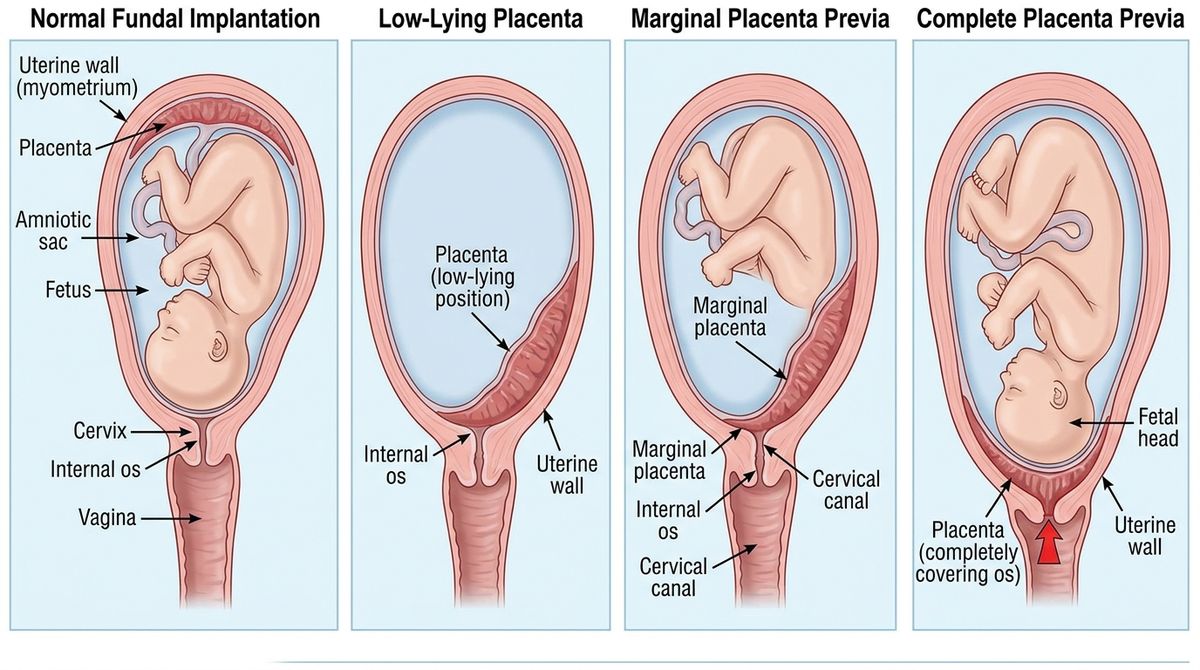
Normal Implantation vs. Low-Lying vs. Previa Types
High-resolution medical diagram contrasting normal fundal attachment against low-lying placenta, marginal previa, and complete previa covering the internal cervical os.
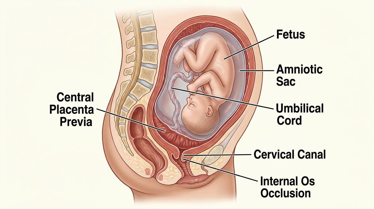
Central Placenta Previa (Complete Previa Totalis)
Detailed sagittal anatomical illustration of central placenta previa, showing the placenta positioned centrally across the lower uterine segment, completely covering the internal cervical os.
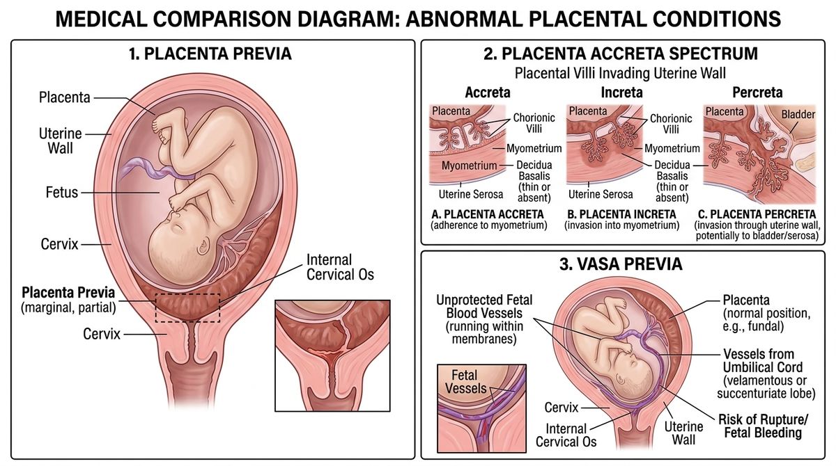
Placenta Previa vs. Accreta Spectrum vs. Vasa Previa
Triple comparative medical illustration showing low placental location (previa), invasive myometrial anchoring (accreta/increta/percreta), and exposed fetal membrane vessels (vasa previa).
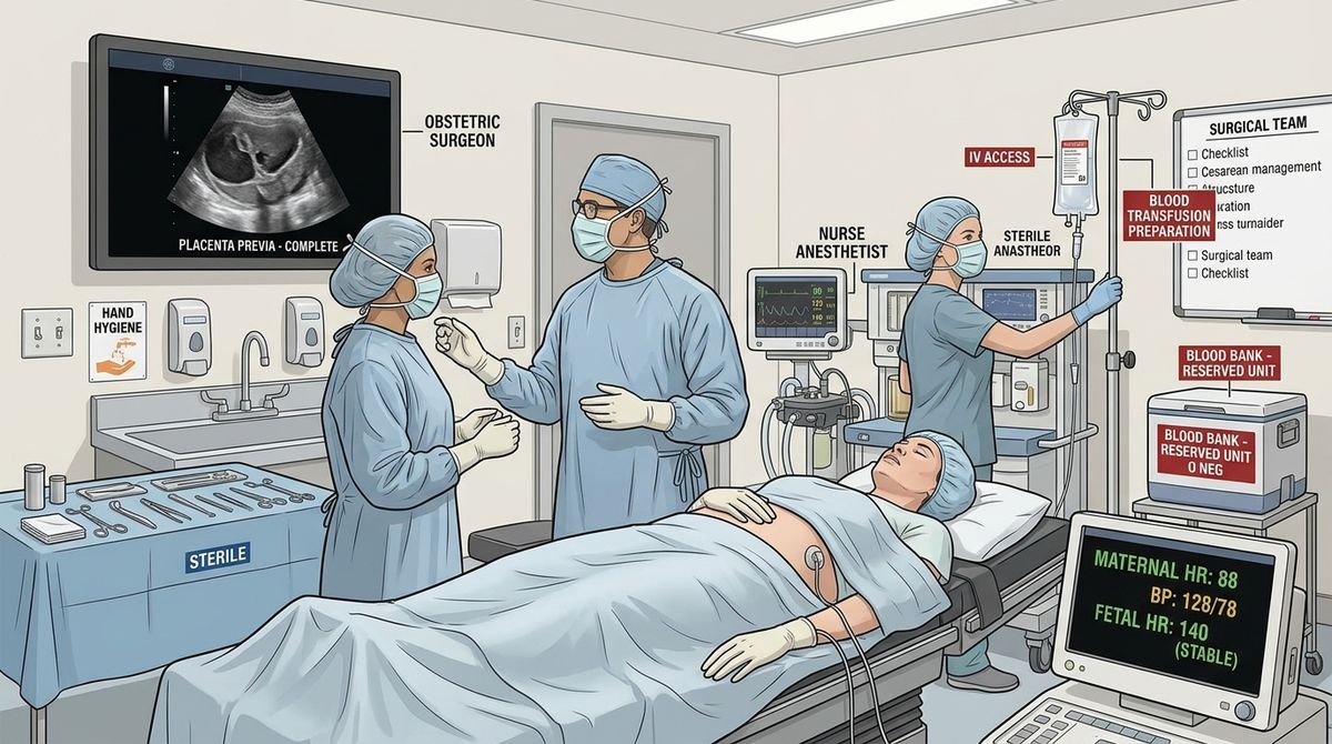
Multidisciplinary Cesarean Suite & Hemorrhage Readiness
Hospital labor and delivery surgical suite prepared for planned cesarean section at 36+0 to 37+6 weeks, continuous vitals, IV access, and blood bank crossmatch reservation.
1. What Happens in Placenta Previa?
Normally, the fertilized blastocyst implants in the upper, thick portion of the uterine wall (fundus or upper corpus), well away from the internal cervical opening. In placenta previa, the placenta attaches in the lower segment of the uterus and either encroaches upon or completely covers the internal opening of the cervix.
Low-Lying Placenta
The placental edge is positioned close to the internal cervical os (typically defined as within 20 mm / 2 cm of the internal os on transvaginal sonography), but does not cover the opening. Many low-lying placentas allow for safe vaginal delivery if the distance exceeds 20 mm near term.
Placenta Previa (Partial / Complete)
Placental tissue directly overlays the internal cervical os, partially or completely. Because vascular placental cotyledons sit immediately above the birth canal, the cervical effacement and dilation that occur in the third trimester shear these fragile attachments, leading to severe hemorrhage.
9 out of 10 Low-Lying Placentas Resolve by the Third Trimester
A placenta that appears low at the routine 18–20 week mid-pregnancy anatomy scan very often does not stay low. As pregnancy advances, the lower uterine segment elongates rapidly from approximately 0.5 cm to over 5 cm in length, and preferential blood supply causes the placenta to grow toward the richly vascular fundus (trophotropism). The Royal College of Obstetricians and Gynaecologists (RCOG) reports that approximately 9 out of 10 women (90%) with a low-lying placenta at 20 weeks no longer have a low-lying placenta at the follow-up 32-week ultrasound.
Summary: Clinical Essentials of Placenta Previa
Placenta previa = low placenta + risk of painless antepartum bleeding + ultrasound diagnosis + careful surveillance + usually planned cesarean if it persists over the cervix.
The major danger is massive obstetric hemorrhage, so any bleeding in the second half of pregnancy should be treated as requiring urgent professional assessment rather than monitored at home.
Placenta Previa: Questions & Clinical Clarifications
Key clinical points frequently encountered in high-risk obstetric consultations.
QCan a low-lying placenta move up later in pregnancy?
Yes, absolutely. The phenomenon often called "placental migration" occurs as the lower uterine segment elongates from 0.5 cm in early pregnancy to over 5 cm in the third trimester. Additionally, the placenta grows preferentially toward the richly vascular fundus (trophotropism). The Royal College of Obstetricians and Gynaecologists (RCOG) reports that approximately 9 out of 10 women (90%) diagnosed with a low-lying placenta at their 20-week anatomy scan no longer have a low-lying placenta at follow-up scans.
QWhy is a digital vaginal or cervical examination contraindicated in placenta previa?
A digital vaginal examination (inserting gloved fingers into the vagina or cervix) is strictly contraindicated in any patient with suspected or confirmed placenta previa. The manual examination can directly palpate and mechanically disrupt the friable placental cotyledons overlying the cervix, instantly triggering massive, life-threatening maternal hemorrhage and fetal compromise. Diagnosis must always be established safely with transvaginal ultrasound first.
QWhat is the optimal timing for planned delivery in stable placenta previa?
According to current Merck professional guidance and major obstetric consensus guidelines (ACOG/RCOG), planned Cesarean delivery for an uncomplicated, asymptomatic placenta previa is typically scheduled between 36+0 and 37+6 weeks of gestation. This balance maximizes fetal lung and organ maturity while minimizing the risk of spontaneous onset of labor and catastrophic antepartum hemorrhage.
QHow does placenta previa differ from placenta accreta spectrum?
Placenta previa refers to an anatomical location problem where the placenta is positioned abnormally low and covers or lies within 2 cm of the internal cervical os. In contrast, Placenta Accreta Spectrum (PAS) is an abnormal attachment or invasive depth problem where chorionic villi penetrate directly into the uterine myometrium. However, patients with placenta previa who have had prior cesarean deliveries are at dramatically increased risk for placenta accreta.
QWhat should I do if I notice bright-red bleeding after 20 weeks?
Any vaginal bleeding in the second half of pregnancy is an obstetric emergency that cannot be diagnosed or safely monitored at home. You should seek immediate emergency medical care or proceed directly to the nearest hospital obstetric triage unit. Do not insert anything into the vagina (no tampons, no intercourse, no exams) and remain resting on your side during transit.
Authoritative Clinical References & Evidence Base
- • Royal College of Obstetricians and Gynaecologists (RCOG): Green-top Guideline No. 27a: Placenta Praevia and Placenta Accreta: Diagnosis and Management.
- • Merck Manual Professional Edition: Placenta Previa (Abnormal Placental Location), current clinical guidelines for planned Cesarean delivery timing (36+0 to 37+6 weeks).
- • American College of Obstetricians and Gynecologists (ACOG): Obstetric Care Consensus No. 7: Placenta Accreta Spectrum & Practice Bulletin No. 204: Fetal Growth Restriction & Antepartum Hemorrhage.
- • Society for Maternal-Fetal Medicine (SMFM): Clinical Guideline #12: Diagnosis and Management of Vasa Previa and Placenta Previa.
The information provided on this website is for educational purposes only and is not intended as a substitute for professional medical advice, diagnosis, or treatment. Always seek the advice of a qualified healthcare provider with any questions you may have regarding a medical condition.