
For general information only. Not a substitute for professional medical advice.
Medi Info Hub Logo

Retrocaval Ureter (Circumcaval Ureter)
Educational content • Medi Info HubUpdated: 10/19/2026
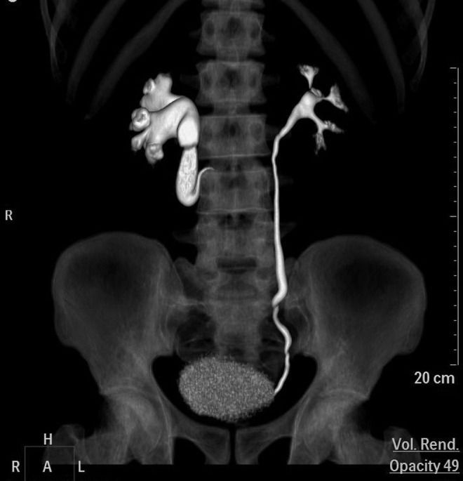
Retrocaval Ureter (Circumcaval Ureter) clinical reference photography
Clinical Photographic Reference
Medical Visual GuideUrology & Congenital Anomalies

Retrocaval Ureter (Circumcaval Ureter)

Educational photographic and clinical reference illustrating presentation, affected anatomy, or clinical evaluation of Retrocaval Ureter (Circumcaval Ureter).

Topic-Accurate VisualClinical Reference

Overview

Retrocaval ureter, also termed circumcaval ureter or preureteral vena cava, is a rare congenital vascular variation in which the right ureter spirals posteriorly around the inferior vena cava (IVC) before emerging anteriorly. This developmental anomaly can cause proximal ureteral compression, hydronephrosis, recurrent flank pain, and urinary tract infections.

What Is It?

Retrocaval ureter occurs due to anomalous persistence of the right posterior cardinal vein during fetal embryogenesis. Instead of coursing straight downward from the kidney to the urinary bladder, the upper third of the right ureter courses medially, passes behind the inferior vena cava, wraps around it, and re-emerges anteromedially before resuming its normal anatomical descent.

Signs & Symptoms

Flank pain (dull aching or intermittent renal colic), microhematuria or gross hematuria, recurrent urinary tract infections, symptoms of nephrolithiasis due to urinary stasis, and palpable right kidney enlargement in advanced hydronephrosis.

Causes

Embryological persistence of the right subcardinal or posterior cardinal veins during the 6th to 8th weeks of gestation. It is almost exclusively right-sided, though exceptionally rare left-sided cases occur in situs inversus or duplicate IVC.

Diagnosis

Contrast-enhanced CT Urography (CTU) is the diagnostic standard of choice, demonstrating the pathognomonic "reverse-J" or "S-shaped" medial deviation of the upper ureter with proximal pelvicalyceal dilation. Magnetic Resonance Urography (MRU) and retrograde pyelography provide additional anatomical confirmation.

Treatment & Management

Asymptomatic patients without significant hydronephrosis or renal compromise are managed conservatively with serial ultrasonography. Symptomatic obstruction requires surgical correction, typically laparoscopic or robotic-assisted pyeloureterostomy or ureteroureterostomy with anterior repositioning of the ureter.

Important: Treatment decisions should always be made in consultation with a qualified healthcare professional. Do not start, stop, or change medications without medical guidance.

Prevention & Risk Reduction

As a congenital anomaly, retrocaval ureter cannot be prevented. Prevention of renal impairment relies on prompt diagnosis and relieving significant urinary outflow obstruction before irreversible cortical thinning occurs.

Living With This Condition

Following surgical correction, most patients achieve complete symptom resolution and normal renal outflow. Long-term follow-up with renal ultrasound and renal function tests confirms lasting relief.

When to Seek Medical Help

When to Contact a Healthcare Professional

Do not delay seeking medical care if needed

Contact a healthcare provider immediately if you experience persistent right-sided flank pain, visible blood in the urine, fever with chills, or symptoms of kidney infection.

Emergency Warning Signs

Frequently Asked Questions

Is retrocaval ureter hereditary?

No, retrocaval ureter is an isolated embryological vascular development variation and is not typically passed down genetically.

Does every patient need surgery?

No. Patients with mild non-progressive hydronephrosis and no flank pain or infections can be monitored conservatively with routine renal scans.

Medical Disclaimer

Medi Info Hub provides general health information for educational purposes only. The information on this website is not medical advice and is not a substitute for diagnosis, treatment, or consultation with a qualified healthcare professional. Do not delay seeking medical care because of information found on this website. In an emergency, contact your local emergency services immediately.

Published by: Medi Info Hub TeamLast updated: October 19, 2026
Retrocaval Ureter — Symptoms, Diagnosis & Treatment | Medi Info Hub | Medi Info Hub