
Breast Cancer

Breast Cancer
Educational photographic and clinical reference illustrating presentation, affected anatomy, or clinical evaluation of Breast Cancer.
Overview: What Is Breast Cancer?
A comprehensive, evidence-based medical guide covering pathophysiology, staging, histopathology, molecular biomarkers, diagnostics, and modern treatment paradigms.
Breast cancer is a cancer that begins when abnormal cells in breast tissue grow and divide without normal control. These abnormal cells can form a tumor and, if they become invasive, can spread into surrounding breast tissue, nearby lymph nodes, or distant organs through the lymphatic system or bloodstream.
Breast cancer can begin in the milk ducts, milk-producing lobules, nipple area, connective tissue, or, rarely, blood and lymphatic vessels. Most breast cancers begin in the ducts or lobules.
Although breast cancer occurs predominantly in women, men can also develop breast cancer because men also have a small amount of breast tissue. Approximately 0.5–1% of breast cancers occur in men.
Breast cancer is not a single disease. Different breast cancers can behave very differently depending on:
Biological Characteristics That Influence Treatment & Prognosis
Rather than a uniform condition, each patient's breast cancer behaves uniquely based on clinical, pathological, and molecular features:
How Does Breast Cancer Develop?
Normal breast cells grow, divide, and die according to tightly controlled biological signals. Cancer develops when genetic changes disrupt these controls. Some changes are inherited from a parent, while most occur during a person's lifetime in individual cells.
Over time, accumulated genetic changes can allow abnormal cells to progress through seven biological steps:
Uncontrolled Growth
Escape normal cell cycle growth arrest signals.
Avoid Normal Cell Death
Evade physiological apoptosis (programmed cell death).
Mass Formation
Proliferate to form a discrete neoplastic tumor mass.
Tissue Invasion
Break through the basement membrane into surrounding stroma.
Vessel Penetration
Intravasate into nearby lymphatic vessels or capillaries.
Lymph Node Seeding
Travel to regional axillary lymph node filter stations.
Distant Organ Metastasis
Colonize distant organs including bone, liver, lungs, or brain.
Breast cancer can therefore exist on a spectrum ranging from noninvasive disease such as ductal carcinoma in situ (DCIS) to invasive cancer and metastatic breast cancer.
What Causes Breast Cancer? & 13 Major Risk Factors
There is usually not one single cause of breast cancer. Breast cancer develops through a combination of genetic, biological, hormonal, environmental, reproductive, and lifestyle factors.
Increasing Age
Age is one of the strongest risk factors. Median age at diagnosis is ~64 years. The largest percentage of diagnoses occurs between ages 65 and 74.
Family History
Having a close relative with breast cancer increases risk. First-degree relatives (mother, sister, daughter) are most important. Family history does not make cancer inevitable.
Inherited Gene Changes
High-penetrance variants substantially increase risk: BRCA1, BRCA2, PALB2, TP53, PTEN, CHEK2, ATM, CDH1, and STK11.
Previous Breast Cancer
Personal history of breast cancer elevates the risk of developing a new primary cancer in the contralateral or same breast.
Prior DCIS or High-Risk Lesions
History of DCIS, atypical ductal hyperplasia (ADH), or atypical lobular hyperplasia (ALH) on prior biopsy increases future breast cancer risk.
Dense Breasts
High fibroglandular breast density increases risk independent of other factors and can mask tumors on 2D screening mammograms.
Previous Radiation Exposure
Mantle or chest wall radiation exposure, particularly during childhood, adolescence, or early adulthood for Hodgkin lymphoma.
Hormonal and Reproductive Factors
Longer lifetime exposure to estrogen: early menarche (before age 12), late menopause (after 55), older age at first full-term pregnancy (>30), or nulliparity.
Menopausal Hormone Therapy (MHT)
Combined estrogen-progestin hormone therapy regimens taken after menopause are associated with an increased risk.
Alcohol Consumption
Alcohol intake is dose-dependently associated with increased breast cancer risk; risk escalates with greater cumulative alcohol exposure.
Physical Inactivity
Sedentary lifestyle and lack of regular moderate-to-vigorous physical activity are consistently associated with higher risk.
Excess Body Weight After Menopause
Overweight and obesity after menopause increase circulating estrogen levels derived from peripheral aromatization in adipose tissue.
Tobacco Exposure
Active cigarette smoking and prolonged heavy secondhand smoke exposure have been associated with increased breast cancer risk.
Breast Cancer by Age & Young Women
Age is useful for understanding population risk, but age alone cannot predict whether an individual will develop breast cancer or survive it. Recent U.S. SEER cancer registry data show the following age distribution at diagnosis:
| Age at Diagnosis | Approx. Percentage of All Diagnoses | Clinical Context & Considerations |
|---|---|---|
| 20–34 | 2.0% | Uncommon; frequently genetic (BRCA) or aggressive biological subtypes |
| 35–44 | 8.6% | Pre-menopausal; higher proportion of triple-negative or HER2+ tumors |
| 45–54 | 17.7% | Perimenopausal transition; USPSTF recommends routine screening at 40 |
| 55–64 | 24.3% | Postmenopausal hormone receptor-positive cancers predominate |
| 65–74 | 27.5% | Highest proportion of all breast cancer diagnoses (peak incidence) |
| 75–84 | 15.1% | High prevalence; treatment tailored to physiological functional status |
| 85+ | 4.9% | Individualized care balancing treatment tolerance and quality of life |
Can Young Women Get Breast Cancer?
Yes. Breast cancer is much less common in younger people, but it can occur before age 40. Some younger patients may have inherited genetic susceptibility, including BRCA1 or BRCA2 variants. Certain aggressive subtypes, including triple-negative breast cancer, are also more frequently diagnosed at younger ages.
Clinical Rule: A new breast lump or other persistent breast change should never be dismissed because of young age.
Important Difference: Risk Factor vs. Cause
Incorrect. Most people diagnosed with breast cancer do not have a known inherited mutation or family history.
Incorrect. Risk factors change statistical probability; they do not dictate an individual's destiny.
Signs, Symptoms & Major Types of Breast Cancer
Recognizing warning signs, distinguishing benign conditions, and understanding histological and molecular classifications.
Breast cancer may cause no symptoms in its early stages. Screening mammograms can detect some cancers before they produce noticeable changes. When symptoms develop, early clinical evaluation is essential.
Breast Lump or Thickening
A new firm lump or thickened area in the breast or armpit that feels distinct from surrounding normal tissue.
Change in Size or Shape
Noticeable progressive asymmetry, swelling, or enlargement of one breast.
Skin Dimpling & Peau d'Orange
Skin puckering, tethering, thickening, or orange-peel texture caused by dermal lymphatic infiltration.
Nipple Inversion & Flattening
New spontaneous retraction, inward pulling, flattening, or deviation of the nipple.
Spontaneous Nipple Discharge
Bloody, clear, or serous discharge from a single duct without squeezing or stimulation.
Persistent Breast or Nipple Pain
Localized, focal discomfort that persists across multiple menstrual cycles and does not resolve.
Armpit or Collarbone Swelling
Enlarged axillary or supraclavicular lymph nodes that feel firm, fixed, or persistently swollen.
Inflammatory Skin Changes
Rapidly spreading erythema, warmth, heaviness, and edema involving more than one-third of the breast.
Nipple Scaling & Crusting
Persistent eczema-like flaking, crusting, itching, or ulceration of the nipple and areola (Paget disease).
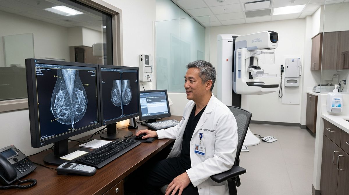
High-resolution digital mammography radiograph illustrating breast fibroglandular density grading, microcalcification clusters, and architectural structural tissue features evaluated in diagnostic oncology screening.
Major Histological Types of Breast Cancer
Breast cancers are classified pathologically according to their specific cellular origin and histological growth pattern:
1. Ductal Carcinoma in Situ (DCIS)
Stage 0 NoninvasiveAbnormal proliferation of cells confined entirely inside milk ducts without breaching the basement membrane into surrounding stroma. If left untreated, some DCIS can progress to invasive cancer.
2. Invasive Ductal Carcinoma (IDC)
70–80% of All CasesThe most common breast cancer. Begins in epithelial cells lining a milk duct, breaks through ductal wall into stroma, with potential to invade lymphatic channels and bloodstream.
3. Invasive Lobular Carcinoma (ILC)
10–15% of CasesArises in milk-producing lobules. Cells grow in single-file discohesive sheets due to E-cadherin loss. Can be harder to detect by palpation or mammogram; higher incidence of bilaterality.
4. Inflammatory Breast Cancer (IBC)
1–5% (Aggressive)Rapidly progressive disease where tumor emboli occlude dermal lymphatic channels. Causes breast erythema, warmth, edema, and peau d'orange, often without a discrete lump.
5. Triple-Negative Breast Cancer (TNBC)
~15% of CancersLacks estrogen receptors (ER-), progesterone receptors (PR-), and HER2 overexpression. Tends to be biologically aggressive with higher recurrence rates; treated with chemo and immunotherapy.
6. HER2-Positive Breast Cancer
15–20% of CancersOverexpresses human epidermal growth factor receptor 2 protein due to gene amplification. Biologically fast-growing, but exceptionally responsive to targeted anti-HER2 monoclonal antibodies.
7. Hormone-Receptor-Positive (HR+)
65–75% of CancersTumors that express estrogen (ER) or progesterone (PR) receptors. Stimulated by circulating hormones and highly responsive to endocrine therapies like tamoxifen and aromatase inhibitors.
8. Paget Disease of the Breast
1–3% of CancersRare cancer involving the nipple and areola, presenting with eczematous crusted changes, discharge, and burning. Frequently associated with underlying DCIS or invasive cancer.
9. Metaplastic Breast Cancer
<1% Rare VariantAggressive subtype where adenocarcinoma cells differentiate into spindle, squamous, or sarcomatous mesenchymal elements; typically triple-negative and chemo-resistant.
10. Phyllodes Tumors
Stromal OriginDevelop in the periductal stromal connective tissue rather than glandular epithelium. Most are benign, but some are borderline or malignant sarcomas requiring wide surgical excision.
11. Lobular Carcinoma in Situ (LCIS)
High-Risk MarkerNot a true cancer or direct precursor. Rather, it represents a strong biological marker of increased bilateral breast cancer risk, managed with close surveillance or chemoprevention.
Intrinsic Molecular Subtypes
In modern oncology, gene expression profiling categorizes breast cancers into intrinsic molecular subtypes that determine biological behavior and therapeutic sensitivity:
Luminal A
Most favorable prognosis
Endocrine therapy; chemo rarely needed
Luminal B
Intermediate prognosis
Endocrine therapy + chemo + HER2 agents
HER2-Enriched
Aggressive; targeted therapy
Anti-HER2 biologics + dual chemotherapy
Basal-Like (TNBC)
Aggressive; earlier recurrence
Chemotherapy + immunotherapy checkpoint
Diagnosis, Pathology, Biomarkers & Staging
From clinical breast exam to digital imaging, image-guided biopsy, receptor biomarkers, and TNM staging.
How Is Breast Cancer Diagnosed? The 6-Step Clinical Process
Comprehensive Medical & Family History
Clinician reviews presenting symptoms, onset timeline, past breast biopsies, radiation exposure, menstrual and reproductive milestones, menopausal hormone use, and family cancer history on maternal and paternal lineages.
Clinical Breast Examination (CBE)
Thorough inspection and manual palpation of breasts, chest wall, axillary lymph node basins, and supraclavicular fossae in upright and supine positions. Clinical examination cannot alone confirm or exclude malignancy.
Digital Mammography (2D & 3D Tomosynthesis)
Standard low-dose X-ray imaging detecting soft-tissue masses, pleomorphic microcalcifications, and architectural distortions. Screening mammograms evaluate asymptomatic women; diagnostic mammograms investigate abnormal findings or focal symptoms.
Targeted Breast Ultrasound
High-frequency sound waves evaluate focal lesions, differentiate fluid-filled simple cysts from solid neoplasms, assess dense breast parenchyma, and guide real-time biopsy needle localization.
Contrast-Enhanced Breast MRI
High-field magnetic resonance imaging with intravenous gadolinium contrast for high-risk surveillance, evaluating disease extent, detecting multifocal or bilateral tumors, and assessing breast implants.
Confirmatory Breast Biopsy
Definitive confirmatory test. While imaging identifies suspicious abnormalities, histopathological biopsy examination is generally required to definitively diagnose breast cancer.
Types of Breast Biopsy
Uses a thin needle to aspirate fluid or loose cells; useful for cystic lesions and palpable axillary lymph nodes.
Uses a hollow needle to harvest tissue cylinders, preserving histology architecture and providing ample tissue for ER, PR, HER2, and Ki-67 testing.
Performed under real-time ultrasound, stereotactic mammography, or MRI guidance to pinpoint nonpalpable calcifications or masses.
Minor operating procedure to remove part or all of a suspicious mass when needle biopsy is discordant or inconclusive.
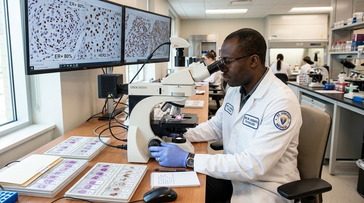
Core needle biopsy histopathological evaluation demonstrating Nottingham histologic grading and immunohistochemistry (IHC) profiling for Estrogen (ER), Progesterone (PR), and HER2/neu receptor status.
Pathology Examination & Crucial Biomarkers
The pathology report guides all systemic therapy choices. Standard immunohistochemical (IHC) profiling tests four vital markers:
Estrogen Receptor (ER)
ER-positive cancer cells are stimulated by estrogen. Treated with endocrine medications (tamoxifen, aromatase inhibitors) to shut down estrogen-driven growth.
Progesterone Receptor (PR)
PR expression confirms an intact, functional estrogen receptor pathway and predicts greater responsiveness to hormone therapies.
HER2 Receptor
HER2 is a cell-surface tyrosine kinase receptor. Cancers with HER2 overexpression/amplification receive targeted monoclonal antibody therapy (trastuzumab, pertuzumab).
Ki-67 Proliferation Index
Nuclear protein measuring percentage of actively dividing tumor cells. High Ki-67 indicates rapidly multiplying cancer cells, aiding chemotherapy decisions.
Genetic Testing & Multigene Tumor Assays
Inherited Germline Genetic Testing
Tests inherited DNA from blood or saliva for hereditary pathogenic cancer variants (BRCA1, BRCA2, PALB2, TP53, PTEN, etc.). Especially recommended for:
- Diagnosis at young age (≤50 years)
- Triple-negative breast cancer at any age ≤60
- Multiple relatives with breast, ovarian, pancreatic, or prostate cancer
- Male breast cancer in family
- Ashkenazi Jewish ancestry
Multigene Genomic Tumor Assays
Different from inherited testing; these tests analyze the tumor itself to predict recurrence risk and chemotherapy benefit in ER+, HER2- early breast cancer:
- Oncotype DX (21-gene recurrence score): Predicts chemotherapy benefit and 10-year distant recurrence risk.
- MammaPrint (70-gene profile): Classifies genomic high vs. low risk of distant metastasis.
- Helps safely spare low-risk patients from toxic chemotherapy.
Breast Cancer Staging: Stage 0 to Stage IV
Staging incorporates the American Joint Committee on Cancer (AJCC) TNM framework combined with tumor grade and ER, PR, HER2 status:
Carcinoma in Situ
DCIS; noninvasive abnormal cells confined inside ducts with zero stromal or nodal spread.
Early Invasive
Tumor ≤2 cm; no lymph node spread or microscopic micrometastases in sentinel node only.
Localized / Regional
Tumor 2–5 cm with 1–3 axillary nodes, or >5 cm without nodal involvement.
Locally Advanced
Extensive regional spread to 4–9+ axillary nodes, internal mammary nodes, chest wall, or skin (inflammatory).
Metastatic Disease
Disseminated cancer spread beyond regional nodes to distant organs (bone, liver, lungs, brain).
What Happens After a Suspicious Mammogram?
A suspicious screening mammogram does not mean cancer. In fact, fewer than 10% of women called back for additional testing have breast cancer. Standard next steps include:
- Diagnostic mammogram spot-compression or magnification views
- Targeted breast ultrasound to evaluate focal densities or cysts
- Short-interval 6-month imaging follow-up for probably benign (BI-RADS 3) findings
- Image-guided needle biopsy for BI-RADS 4 or 5 suspicious lesions
How Breast Cancer Is Treated
Evidence-based multimodal therapies: surgery, radiation, chemotherapy, targeted biologics, and metastatic organ-specific management.
Most breast cancer patients receive a combination of treatments tailored to their specific tumor stage, biomarker profile, and personal preferences rather than any single intervention alone.
Surgical Resection
Local ControlBreast-conserving surgery (lumpectomy) with clear margins followed by radiation, or total mastectomy. Always paired with sentinel lymph node biopsy to evaluate nodal staging without unnecessary axillary dissection.
Radiation Therapy
Locoregional ControlHigh-energy X-rays destroy residual microscopic cancer cells in the breast, chest wall, or regional nodal beds, cutting recurrence rates in half following breast-conserving surgery.
Chemotherapy
Systemic EradicationCytotoxic agents destroying rapidly dividing cells. Delivered as neoadjuvant therapy (before surgery to shrink large tumors) or adjuvant therapy (after surgery to prevent distant recurrence).
Endocrine (Hormone) Therapy
ER+ Cancers (5–10 Years)Blocks estrogen receptors (Tamoxifen in premenopausal women) or prevents estrogen production (Aromatase Inhibitors: anastrozole, letrozole, exemestane in postmenopausal women).
HER2-Targeted Biologics
HER2+ CancersMonoclonal antibodies (trastuzumab, pertuzumab) and revolutionary antibody-drug conjugates (T-DM1, trastuzumab deruxtecan) targeting HER2 oncoproteins.
Immunotherapy Checkpoints
Triple-Negative CancersImmune checkpoint inhibitors (such as pembrolizumab) combined with chemotherapy for high-risk early or metastatic PD-L1-positive triple-negative breast cancer.
Targeted Inhibitors (CDK4/6 & PARP)
Genomic PrecisionCDK4/6 inhibitors (palbociclib, ribociclib, abemaciclib) for metastatic HR+ cancer; PARP inhibitors (olaparib, talazoparib) for patients with inherited BRCA mutations.
Sentinel Lymph Node Surgery
Lymphatic StagingInjecting radioactive tracer or blue dye identifies the first 1–3 filter nodes receiving breast drainage, preventing lymphedema caused by full axillary lymph node dissection.
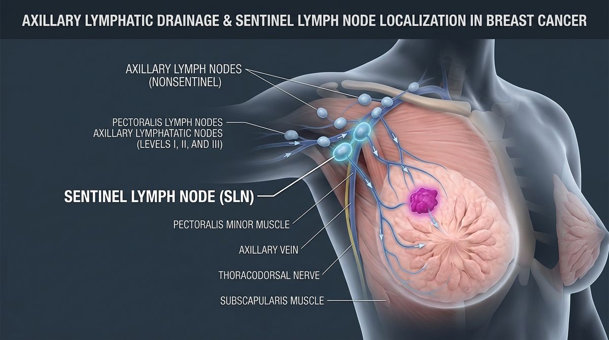
Regional lymphatic drainage anatomy illustrating Level I, II, and III axillary lymph node stations and sentinel lymph node mapping for accurate pathological tumor staging without routine full axillary dissection.
Metastatic Breast Cancer & Organ-Specific Care
Metastatic breast cancer (Stage IV) occurs when cancer cells spread through blood or lymph vessels to distant organs. Common metastatic sites include:
Bone Metastasis
Signs: Bone pain, pathological fractures, elevated blood calcium (hypercalcemia).
Management: Bone-modifying agents (zoledronic acid, denosumab), palliative radiation, orthopedic stabilization.
Liver Metastasis
Signs: Abdominal fullness, fatigue, elevated liver enzymes (LFTs), jaundice in advanced cases.
Management: Systemic targeted and chemotherapy regimens, liver-directed therapies, monitoring hepatic function.
Lung Metastasis
Signs: Shortness of breath, persistent cough, chest discomfort, pleural effusion.
Management: Systemic systemic treatment, thoracentesis for symptomatic pleural fluid, supplemental oxygen.
Brain Metastasis
Signs: Headaches, focal weakness, seizures, balance changes, cognitive shifts.
Management: Stereotactic radiosurgery (SRS), whole-brain radiation, brain-penetrating targeted biologics (tucatinib).
Survival Statistics, Prevention & Screening Guidelines
SEER survival data across stages and subtypes, lifestyle risk reduction, high-risk prevention strategies, and screening recommendations.
U.S. SEER 5-Year Relative Survival Statistics
A survival percentage describes outcomes observed in populations diagnosed during earlier surveillance periods; it is not a prediction for an individual patient. Recent National Cancer Institute (NCI) SEER data reported by the American Cancer Society document the decisive impact of early diagnosis:
| Extent of Disease at Diagnosis | 5-Year Relative Survival Rate | Clinical Anatomical Definition |
|---|---|---|
| Localized Disease | >99% (99.3%) | Cancer is confined strictly inside the breast; no nodal or distant spread |
| Regional Disease | 87.0% (86.3%) | Spread to nearby regional axillary lymph nodes or chest structures |
| Distant (Metastatic) Disease | 32.0% (31.0%) | Cancer has spread to distant organs (bone, liver, lungs, brain) |
| All SEER Stages Combined | 92.0% | Combined overall 5-year relative survival across all female breast cancers |
Survival According to Molecular Subtype
Localized TNBC: 91% • Regional TNBC: 66% • Distant TNBC: 12%
Regional IBC: 53% • Distant IBC: 22%
Can Breast Cancer Be Prevented? Evidence-Based Risk Reduction
While no method can guarantee prevention, evidence demonstrates several modifiable behaviors that lower individual risk:
Regular Physical Activity
Engage in at least 150 minutes of moderate or 75 minutes of vigorous aerobic exercise weekly to regulate insulin and estrogen metabolism.
Healthy Postmenopausal Weight
Prevent excess adipose tissue accumulation after menopause, minimizing peripheral aromatization into circulating estrogens.
Avoid or Limit Alcohol
Alcohol increases circulating estrogen levels and cellular oxidative stress; limiting or avoiding alcohol directly reduces risk.
Breastfeeding When Feasible
Lactation for several months after childbirth reduces lifetime ovulatory cycles and promotes full terminal differentiation of breast cells.
Know Family Cancer History
Track both maternal and paternal diagnoses to enable early genetic assessment and personalized surveillance protocols.
Judicious Hormone Therapy
Carefully discuss the benefits and risks of combined estrogen-progestin therapy with your physician, opting for lowest dose/duration when needed.
Strategies for People at Very High Genetic Risk
Patients with high-penetrance pathogenic variants (BRCA1, BRCA2, PALB2, TP53) or extreme familial clustering may consider specialized risk-reducing strategies:
Alternating annual breast MRI and digital mammography every 6 months starting at age 25–30.
Selective estrogen receptor modulators (Tamoxifen, Raloxifene) or aromatase inhibitors cutting risk by up to 50%.
Bilateral prophylactic mastectomy reduces risk by >90%; bilateral salpingo-oophorectomy cuts ovarian cancer risk.
Screening Guidelines (USPSTF & ACS)
The U.S. Preventive Services Task Force recommends biennial screening mammography from ages 40 through 74 for women at average risk. High-risk women require earlier and supplemental MRI screening.
Breast Self-Awareness
Be familiar with the normal appearance and feel of your breasts. Promptly report any new or persistent lump, nipple inversion, discharge, or skin dimpling to your healthcare provider.
When to Seek Help, Questions for Doctors & Key Takeaways
Emergency warning signs, red-flag symptoms checklist, essential doctor questions, and clinical takeaways.
Prompt medical consultation is strongly advised for any new or persistent change:
Never attempt to self-diagnose breast changes from internet articles; clinical examination and diagnostic imaging are necessary.
Questions to Ask Your Medical Oncologist & Surgical Team
Summary: What You Need to Know
- •Breast cancer is a group of biologically distinct diseases rather than one single condition.
- •Increasing age is the strongest non-modifiable risk factor; median age at diagnosis is 64 years.
- •Young women can still develop breast cancer, often presenting with genetic (BRCA) or aggressive subtypes.
- •Regular mammography screening detects breast cancer early, when localized 5-year survival exceeds 99%.
- •A tissue biopsy is required for definitive confirmation, histological grading, and biomarker typing.
- •ER, PR, HER2, and genomic assays dictate personalized multimodal therapy (surgery, radiation, chemo, endocrine, and biologics).
- •Physical activity, maintaining healthy weight after menopause, and limiting alcohol reduce individual risk.
- •A suspicious breast change does not automatically mean cancer, but all persistent changes warrant prompt evaluation.
Sources & Medically Recognized References
This clinical guide was compiled using data and guidelines from premier international oncology organizations:
Surveillance, Epidemiology, and End Results (SEER) Program: Female Breast Cancer Diagnostic Age Distribution & 5-Year Relative Survival Statistics.
Breast Cancer Facts & Figures: Staging, Biomarkers (ER/PR/HER2), and Population Survival Rates.
Breast Cancer Global Fact Sheets: Etiology, Risk Factors, and Global Disease Burden.
Screening for Breast Cancer: Recommended Biennial Screening Mammography Ages 40–74.
Medical Disclaimer
Medi Info Hub provides general health information for educational purposes only. The information on this website is not medical advice and is not a substitute for diagnosis, treatment, or consultation with a qualified healthcare professional. Do not delay seeking medical care because of information found on this website. In an emergency, contact your local emergency services immediately.